
By Valerie Lapointe
Ultrasound imaging, a mainstay of medical diagnostic testing, produces dramatically sharper pictures today than when it was introduced more than 50 years ago.
Yet the training procedures used to master the technology haven’t evolved much in that time. A new ultrasound training device, called Dr. Doppler, developed by Illinois-based SIMnext, aims to change that.
Dr. Doppler made its debut during a press event at the MATTER health care incubator in the Merchandise Mart in Chicago this month. This week, the team behind it demonstrated the device at the International Meeting on Simulation in Healthcare, a healthcare simulation conference featuring peer-reviewed learning and the latest industry-specific products in San Diego, Ca.
“The machine gives you the ability to see all types of ailments in simulation, instead of waiting for a patient with the condition to show up.” said Paul Pribaz, executive director at SIMnext. “patients shouldn’t bear the brunt of medical training and Dr. Doppler removes some of that.”
Ultrasound imaging, most commonly known for its use in prenatal care, can also be used to visualize muscles, tendons, internal organs, and major blood vessels, giving it a range of applications across multiple medical disciplines. Ultrasound technology is also portable, and substantially lower in cost when compared to a CT or x-ray exam, which require potentially harmful low-level radiation.

However, in order to make use of ultrasound technology, patients often have to travel to a hospital and work with a technician who typically has 2-4 years of specialized training. During that training, technicians complete several credits of classroom instruction before moving on to working in a clinical setting, first using healthy volunteers and then real patients.
Dr. Doppler, a portable ultrasound simulation training system, offers a bridge-gap between classroom instruction and live tissue. Able to simulate dozens of diagnostic scenarios rarely seen in a clinical setting, it also provides targeted performance feedback through its accompanying mobile app.
“There are currently no other simulation tools that allow you to look at live blood flow through a vessel, you have to learn on a real patient with a known pathology, and even then, you have a limited number of learners that can practice on any given patient.” said Dr. David Salzman, director of simulation for undergraduate education at Northwestern University. “This allows a teacher to control the conditions of the pathology presented, and allow students to practice until they are competent. Nothing proceeds this in the marketplace.”rr
While ultrasound technicians are trained to work with any part of the body, the projected application of this device is to provide specific body part training to practicing general physicians and specialists.
“Ultrasound techs go to several years of school to achieve proficiency, but any doctor can spend a little time using this machine and gain mild proficiency on one part of the body.” said Dr. Thomas Cusack, clinical professor of radiology at the University of Illinois.
The idea is to give physicians expertise to interpret an ultrasound and diagnose a patient without sending them to a hospital, keeping cost, and number of appointments down for the patient as well as incorporating ultrasound into everyday medical practice. “Our goal here is to keep patients out of hospitals and in doing so, allow clinicians to work to the full extent of their license,” added Pribaz.
“If a cohort of doctors can be taught to do one specific procedure, even if that is just identifying what is normal and abnormal, it has the potential to eliminate unnecessary diagnostic tests, save money, and reduce the backlog of testing a hospital regularly experiences,” said Salzman.
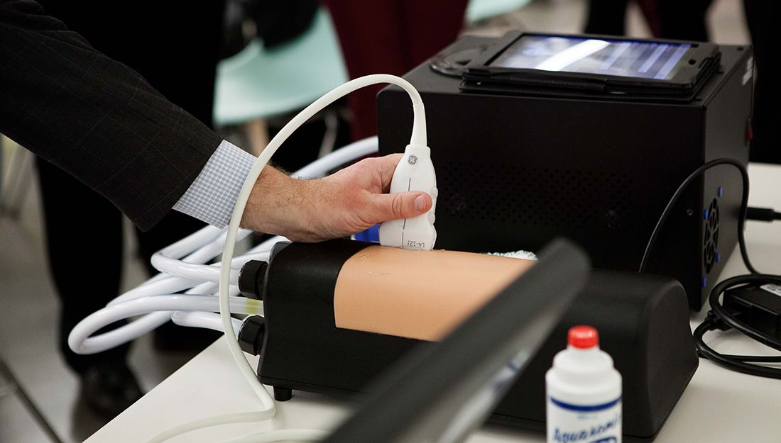
The currently available module is a core competency trainer that allows medical personnel to practice basic hydraulic principles, hand-eye coordination, and methods of diagnosis. While on the surface it only looks like a black box with a squishy beige covering over the center, the strength of the technology lies below the surface.
A specially formulated fluid, designed to mimic the viscosity of blood, is pumped into the module based on the type of simulation selected. The fluid can simulate blood flow patterns associated with a specific pathology or condition.
According to an instructional video on the SIMnext website, the wand passes over an emulator, the flesh-like surface that works with the mobile app that houses the custom training modules. Everything is synchronized via wireless communication, allowing clinicians to observe, interpret, and interact with pathologies in an environment identical to what they will experience working with live patients.” The program can also simulate a variety of normal and abnormal outcomes, with module programs based on real human data.
The app accompanying the device not only houses the training modules, but also compiles data all those who use it. “I’m able to get data on 800 nurses in less than an hour, and from that determine which nurses need more training, and which don’t, saving time and money for the hospital and it’s staff” said registered nurse and clinical education programs specialist, Nikki Delinski.
Only the core competency module is currently available, but four other anatomical phantoms are to be released in 2016-2017, which will improve training on carotid, femoral, testicular, and cerebral areas of the body.
SIMnext is headquartered in Peoria, Illinois, and recently partnered with Limbs & Things, headquartered in Bristol, United Kingdom, to distribute Dr. Doppler. “We are very excited to be working with the Limbs & Things team on the launch of Dr. Doppler,” said Pribaz. “The launch of this system completely changes the way clinicians develop their skill set and diagnostic abilities using ultrasound.”
The new partnership will make the DR Doppler system available in eight countries beginning in the first quarter of 2016. It is projected that the device also might help train doctors in a third world health care context, as adequate health care training is often lacking in developing nations.